45 ear diagram without labels
Ear Anatomy: Understanding the Outer, Middle, and Inner Parts of the Ear The external auditory meatus, or ear canal, is a narrow canal that leads from the concha to the tympanic membrane, or eardrum. Sound waves are delivered through this canal. This canal is prone to ear infections. Tragus This is the small, rigid part of the ears along the front of the ear, adjacent to the face. Ear Diagram Quiz - ProProfs Settings Create your own Quiz . Questions and Answers 1. What is number 1? A. Auricle B. Auditory Tube C. Pharynx D. Stapes 2. What is number 2? A. Incus B. Semicircular Canals C. Cochlea D. Eardrum 3. What is 3? A. Incus B. Auditory Tube C. Oval Window D. Malleus 4. What is 4? A. Semicircular canals B. External acoustic meatus C. Cochlea D.
Human Ear Diagram Photos and Premium High Res Pictures - Getty Images Browse 240 human ear diagram stock photos and images available, or start a new search to explore more stock photos and images. Atrial fibrillation, supraventricular arrhythmia in the heart a supraventricular arrhythmia. In the design of a heart seen in section, the stasis of... The Gallbladder And Bile Ducts In Situ.

Ear diagram without labels
Unit 3: Sensation and Perception - AP Psychology The Five Senses. Eye Diagram Without Labels. Eye Diagram With Labels. Retina (Rods and Cones) Diagram. Ear Diagram Without Labels. Ear Diagram With Labels. The Middle and Inner Ear Diagram. Perception - Grouping. Optical Illusions. Human Ear: Structure and Functions (With Diagram) The sound waves are collected by the external ear up to some extent. They pass through the external auditory meatus to the tympanic membrane which is caused to vibrate. The vibrations are transmitted across the middle ear by the malleus, incus and to the stapes bones. The latter fits into the fenestra ovalis. Ear Anatomy - Outer Ear | McGovern Medical School Ear Anatomy - Outer Ear. The outer ear comes in all types of shapes and sizes. This structure helps to give each of us our unique appearance. The medical term for the outer ear is the auricle or pinna. The outer ear is made up of cartilage and skin. There are three different parts to the outer ear; the tragus, helix and the lobule.
Ear diagram without labels. Image result for ear structure without label | Ear diagram, Ear anatomy ... Parts of the Eye Diagram for 4th graders | Lesson 2 Grade 3 - Grade 4 Activities. The olfactory system enables us to detect odors. Our sense of smell involves nerves, the brain, and sensory organs such as the nose and olfactory bulbs. Original Dust Brush is a good, comfortable, fast - to - use product. Ear Diagram Photos and Premium High Res Pictures - Getty Images ear diagram sound dog ear diagram 250 Ear Diagram Premium High Res Photos Browse 250 ear diagram stock photos and images available, or search for inner ear diagram or human ear diagram to find more great stock photos and pictures. of 5 NEXT Ear Diagram and Labeling Worksheet - twinkl.com The second page shows an ear diagram without labels. The final page shows the labels linking to the beginning letters of each feature, but without the words list. With this Parts of the Ear Labelling Activity, you can test children's memory and their ability to connect different visual signs with words and letters. Anatomy Human Ear Diagram Worksheet 12 Images of Anatomy Human Ear Diagram Worksheet. Blank Ear Diagram. Human Eye Diagram Unlabeled. General and Special Senses Worksheet. Male and Female Reproductive System Functions. Skeletal System Coloring Pages. Label the Parts of the Heart Worksheet. Parts of the Human Respiratory System. Parts of the Human Respiratory System.
Human Ear Diagram - Bodytomy Helix: It is the prominent outer rim of the external ear. Antihelix: It is the cartilage curve that is situated parallel to the helix. Crus of the Helix: It is the landmark of the outer ear, situated right above the pointy protrusion known as the tragus. Auditory Ossicles: The three small bones in the middle ear, called malleus, stapes, and incus, are connected. Well-Labelled Diagram Of Ear With Explanation - BYJUS A brief description of the human ear along with a well-labelled diagram is given below for reference. Well-Labelled Diagram of Ear The External ear or the outer ear consists of: Pinna/auricle is the outermost section of the ear. The external auditory canal links the exterior ear to the inner or the middle ear. Label the Eye Diagram - Enchanted Learning Label the Eye Diagram. Human Anatomy. Read the definitions, then label the eye anatomy diagram below. Cornea - the clear, dome-shaped tissue covering the front of the eye. Iris - the colored part of the eye - it controls the amount of light that enters the eye by changing the size of the pupil. Lens - a crystalline structure located just behind ... Human Body Parts Images Without Labels - Free Vector Download 2020 Human ear diagram with labels and label of anatomy labeling the ear purposegames nose diagram with label diagrams all labels human ear the ear diagram without labels anatomy human charts. Illustration Of Body Parts Labels It is certainly the most widely studied structure the world over. Human body parts images without labels.
Blank ear diagrams and quizzes: The fastest way to learn - Kenhub Ear diagrams (labeled and unlabeled) Accelerate your learning with interactive quizzes Sources + Show all Ear anatomy overview Although it's not obvious to look at, the ear is anatomically divided into three portions: External (outer) ear Middle ear Inner ear As you can imagine, there's a lot of associated anatomy to learn for each portion! Ear, middle ear, cochlea, | Cochlea Diagram of the three parts of the ear : external ear (E), middle ear (M) and inner ear (I) The outer or external ear (e blue) is composed of the pinna (the visible part!) and the ear canal. The latter is closed off by the eardrum. In the middle ear (m orange), the eardrum is mechanically linked by a chain of three tiny bones (the ossicles) to ... Ear Diagram Unlabeled Best Unlabeled diagram human ear free vector download for commercial use in ai, eps, cdr, svg vector illustration graphic art design format. unlabeled. Test students' knowledge of the human eye and ear as they color and label these diagrams. Human Heart Diagram Labeled - Science Trends Without the heart, the tissues couldn't get the oxygen they need and would die. Along with lymphatic vessels, the blood, blood vessels, and lymph, the heart composes the circulatory system of the body. Let's examine the anatomy of the heart along with some diagrams that show how the heart operates. Anatomy Of The Heart
Structure and Functions of the Ear Explicated With Diagrams Structure and Functions of the Ear Explicated With Diagrams. The ear is another extraordinary organ of the house of wonders, that is, the human body. The ear catches sound waves and converts it into impulses, that the brain interprets, making it understandable and helps the human body differentiate between different sounds.
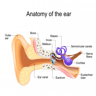
734 Ear Diagram Stock Photos and Images - 123RF diagram of the anatomy of the human ear. Three ossicles: malleus, incus, and stapes (hammer, anvil, and stirrup). The ossicles directly couple sound energy from the ear drum to the oval window of the cochlea. Detailed illustration for educational, medical, biological, and scientific use human ear anatomy, hearing system on white background
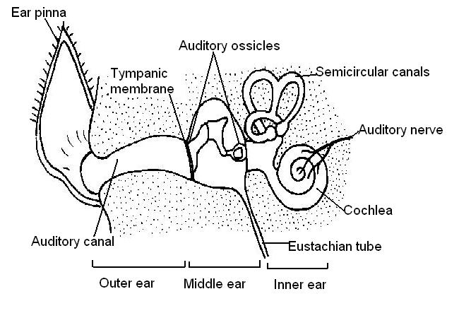
Human Ear Anatomy - Parts of Ear Structure, Diagram and Ear Problems The external (outer) ear consists of the auricle, external auditory canal, and eardrum (Figure 1 and 2). The auricle or pinna is a flap of elastic cartilage shaped like the flared end of a trumpet and covered by skin. The rim of the auricle is the helix; the inferior portion is the lobule. Ligaments and muscles attach the auricle to the head.
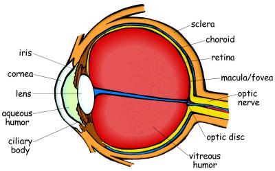
39 diagram of the human eye without labels Diagram of the human eye without labels. Human Ear Diagram - Bodytomy Look no further, this Bodytomy article gives you a labeled human ear diagram and also explains the functions of its different components. The human body is like a big machine, and various processes take place inside it.
25 Heart Diagram Worksheet Blank | Softball Wristband Template ... Nov 16, 2021 - Heart Diagram Worksheet Blank - 25 Heart Diagram Worksheet Blank , Flower Diagram without Labels - thefrangipanitree
Anatomy of the eye: Quizzes and diagrams - Kenhub Take a look at the diagram of the eyeball above. Here you can see all of the main structures in this area. Spend some time reviewing the name and location of each one, then try to label the eye yourself - without peeking! - using the eye diagram (blank) below. Unlabeled diagram of the eye. Click below to download our free unlabeled diagram of ...
Picture of the Ear: Ear Conditions and Treatments - WebMD The fluid-filled semicircular canals (labyrinth) attach to the cochlea and nerves in the inner ear. They send information on balance and head position to the brain. The eustachian (auditory) tube...
Label Parts of the Human Ear - University of Dayton Label Parts of the Human Ear. Select One Auditory Canal Cochlea Cochlear Nerve Eustachian Tube Incus Malleus Oval Window Pinna Round Window Semicircular Canals Stapes Tympanic Membrane Vestibular Nerve. Select One Auditory Canal Cochlea Cochlear Nerve Eustachian Tube Incus Malleus Oval Window Pinna Round Window Semicircular Canals Stapes ...
Ear Anatomy - Outer Ear | McGovern Medical School Ear Anatomy - Outer Ear. The outer ear comes in all types of shapes and sizes. This structure helps to give each of us our unique appearance. The medical term for the outer ear is the auricle or pinna. The outer ear is made up of cartilage and skin. There are three different parts to the outer ear; the tragus, helix and the lobule.
Human Ear: Structure and Functions (With Diagram) The sound waves are collected by the external ear up to some extent. They pass through the external auditory meatus to the tympanic membrane which is caused to vibrate. The vibrations are transmitted across the middle ear by the malleus, incus and to the stapes bones. The latter fits into the fenestra ovalis.
Unit 3: Sensation and Perception - AP Psychology The Five Senses. Eye Diagram Without Labels. Eye Diagram With Labels. Retina (Rods and Cones) Diagram. Ear Diagram Without Labels. Ear Diagram With Labels. The Middle and Inner Ear Diagram. Perception - Grouping. Optical Illusions.


Post a Comment for "45 ear diagram without labels"