44 heart structure with labels
Heart Diagram with Labels and Detailed Explanation - Collegedunia The heart is located under the ribcage, between the lungs and above the diaphragm. It weighs about 10.5 ounces and is cone shaped in structure. It consists of the following parts: Heart Detailed Diagram Heart - Chambers There are four chambers of the heart . The upper two chambers are the auricles and the lower two are called ventricles. How to Draw the Internal Structure of the Heart (with Pictures) - wikiHow Make sure to label the following: Superior Vena Cava Inferior Vena Cava Pulmonary Artery Pulmonary Veins Left Ventricle Right Ventricle Left Atrium Right Atrium Mitral Valves Aortic Valves Aorta Pulmonic Valve (Optional) Tricuspid Valve (Optional) 6 To finish, label "The Human Heart" above the sketch. Tips Use pencil
Fellow of the American Heart Association (FAHA) For those who qualify, election as a Fellow of the American Heart Association recognizes your scientific and professional accomplishments, volunteer leadership and service. By earning the right to include the initials FAHA among your credentials, you let colleagues and patients know that you have been welcomed into one of the world’s most eminent organizations of …

Heart structure with labels
Heart anatomy: Structure, valves, coronary vessels | Kenhub Inside, the heart is divided into four heart chambers: two atria (right and left) and two ventricles (right and left). Diagrams, quizzes and worksheets of the heart | Kenhub Labeled heart diagrams Take a look at our labeled heart diagrams (see below) to get an overview of all of the parts of the heart. Once you're feeling confident, you can test yourself using the unlabeled diagrams of the parts of the heart below. Labeled heart diagram showing the heart from anterior Unlabeled heart diagrams (free download!) Simple heart diagram | Simple heart diagram labeled | Human heart ... Internal structure of human heart shows four chambers viz. two atria and two ventricles and couple of blood vessels opening into them. The wall of two ventricles are strong and sturdy when compared to atria. Before we start, we shall recall the basic proportions of heart and its chambers. The right Auricle is larger than left.
Heart structure with labels. How to draw internal structure of Human heart - Easy version Internal structure of human heart shows four chambers viz. two atria and two ventricles and couple of blood vessels opening into them. The wall of two ventricles are strong and sturdy when compared to atria. Before we start, we shall recall the basic proportions of heart and its chambers. The right Auricle is larger than left. Heart (Human Anatomy): Overview, Function & Structure | Biology The heart is a muscular organ that pumps blood throughout the body. It is located in the middle cavity of the chest, between the lungs. In most people, the heart is located on the left side of the chest, beneath the breastbone. The heart is composed of smooth muscle. It has four chambers which contract in a specific order, allowing the human ... Chapter 19: The Heart Flashcards | Quizlet •Allows heart to beat without friction, gives it room to expand and resists excessive expansion •Parietal pericardium-tough outer, fibrous layer of connective tissue-inner serous layer •Visceral pericardium (a.k.a. epicardium of heart wall)-serous lining of sac turns inward at base of heart to cover the heart surface Eating for a healthy heart - Baker This may help to improve your overall heart health. Unhealthy ‘saturated and trans’ fats should be limited. This fact sheet will help you with: choosing healthy fats and sources; practical ways to include this into your diet; reducing unhealthy fats in your diet; how to read nutrition food labels. Download fact sheet
Carbohydrates | American Heart Association 16/04/2018 · Carbohydrates are either called simple or complex, depending on the food’s chemical structure and how quickly the sugar is digested and absorbed. The type of carbohydrates that you eat makes a difference – Foods that contain high amounts of simple sugars, especially fructose raise triglyceride levels. Triglycerides (or blood fats) are an … The Anatomy of the Heart, Its Structures, and Functions - ThoughtCo The heart is the organ that helps supply blood and oxygen to all parts of the body. It is divided by a partition (or septum) into two halves. The halves are, in turn, divided into four chambers. The heart is situated within the chest cavity and surrounded by a fluid-filled sac called the pericardium. This amazing muscle produces electrical ... Structure of the Heart | The Franklin Institute The heart consists of four chambers: two atria on the top and two ventricles on the bottom. Looking at the Valentine's Day heart, the two rounded humps at the top are rounded like the top of a lower-case "a." The bottom is shaped like a "v." Feel it working What else is inside your heart? heart structure 3D realistic interactive virtual heart structure A common misconception is that all veins carry deoxygenated blood. It is more appropriate to classify veins as vessels carrying blood to the heart. MRI anatomy of the Horse heart The three-dimensional horse and heart on the left can be rotated, zoomed, and panned by clicking and moving the mouse as indicated.
Eplerenone: a blood pressure medicine used to treat heart failure It’s used to treat heart failure and reduce the risk of you having other heart problems or a stroke. It also helps to stop heart failure getting worse. It can sometimes be used to treat a condition called hyperaldosteronism. This is when your body makes too much aldosterone, a hormone that controls your blood pressure. Eplerenone comes as tablets and is only available on … Label the HEART | Circulatory System Quiz - Quizizz True or False: Blood flows in the following sequence in the heart: Vena cava, right atrium, right ventricle, pulmonary artery, lungs, pulmonary veins, left atrium, left ventricle, aorta. Q. True or False: There are four chambers in the heart. Q. Place the pathway of blood through the heart in the correct sequence. Q. Internal Structure of the Heart | Contemporary Health Issues Internal Structure of the Heart. Recall that the heart's contraction cycle follows a dual pattern of circulation—the pulmonary (lungs)and systemic (body) circuits—because of the pairs of chambers that pump blood into the circulation. In order to develop a more precise understanding of cardiac function, it is first necessary to explore the ... heart | Structure, Function, Diagram, Anatomy, & Facts A thin layer of tissue, the pericardium, covers the outside, and another layer, the endocardium, lines the inside. The heart cavity is divided down the middle into a right and a left heart, which in turn are subdivided into two chambers. The upper chamber is called an atrium (or auricle), and the lower chamber is called a ventricle.
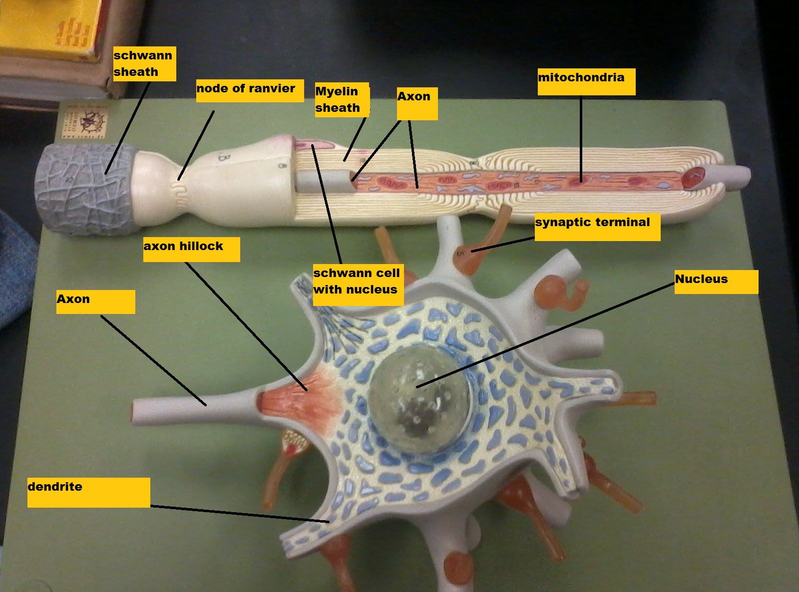
Chapter 14 Nervous Tissue - Biology 4 Human AnatomyProfessor Julie GallagherBarstow Community ...
Heart Anatomy: Labeled Diagram, Structures, Blood Flow ... - EZmed There are 4 chambers, labeled 1-4 on the diagram below. To help simplify things, we can convert the heart into a square. We will then divide that square into 4 different boxes which will represent the 4 chambers of the heart. The boxes are numbered to correlate with the labeled chambers on the cartoon diagram.
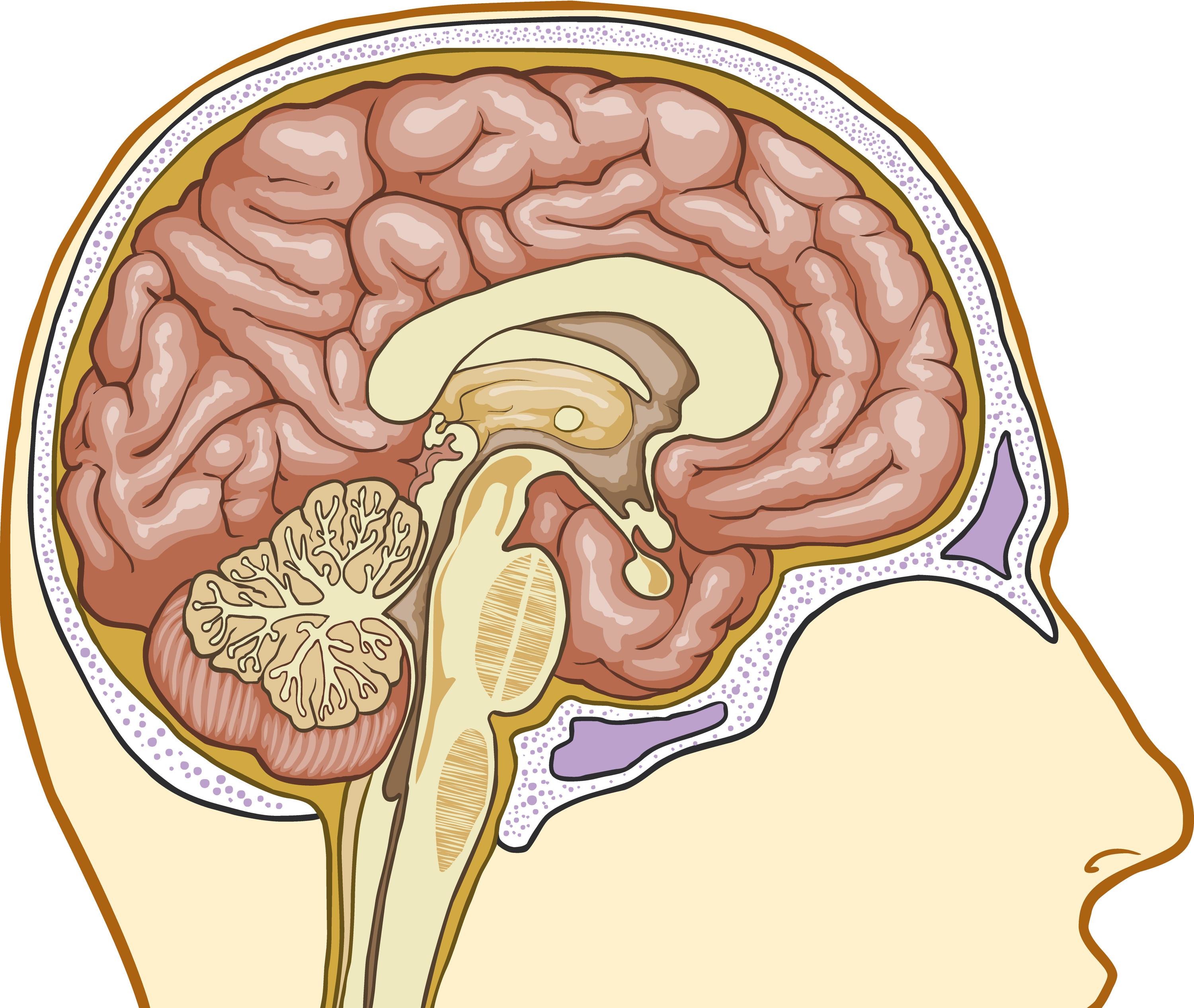
Basic Structure Of The Thalamus - Interactive Biology, with Leslie Samuel
PDF HEART - STRUCTURE - BiologyMad HEART - STRUCTURE • 4 sections Left atrium Right atrium Left ventricle Right ventricle • heart ry artery Pulmonary vein EAS the blood from he left hand side has to be pumped all around the body. • 2 lo heart Atrioventricular valves - between the atrium and the ventricles Semi-lunar valves - in the pulmonary artery and the aorta
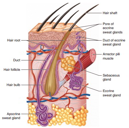
Epidermis And Accessory Structure Formed By The Epidermis And Their Functions
Human Heart - Anatomy, Functions and Facts about Heart - BYJUS To know more about the human heart structure and function, or any other related concepts such as arteries and veins, ... Practice your understanding of the heart structure. Drag and drop the correct labels to the boxes with the matching, highlighted structures. Instructions to use: Hover the mouse over one of the empty boxes. One part in the image gets highlighted. Identify the …
Post a Comment for "44 heart structure with labels"